
Exercise 9 the appendicular skeleton – Exercise 9: The Appendicular Skeleton embarks on an enlightening journey into the intricate world of bones that form our limbs and girdles. This comprehensive guide delves into the structure, function, and clinical significance of this fascinating skeletal system, unveiling its remarkable role in human movement and overall well-being.
The appendicular skeleton, comprising the bones of the upper and lower limbs, pectoral girdle, and pelvic girdle, plays a pivotal role in supporting and protecting our bodies while facilitating a wide range of movements. From reaching for the stars to taking a leisurely stroll, the appendicular skeleton empowers us with the ability to navigate our surroundings with grace and agility.
The Appendicular Skeleton
The appendicular skeleton is the part of the skeleton that consists of the bones of the limbs and the girdles that connect them to the axial skeleton. It is divided into two parts: the upper limb and the lower limb.The
upper limb consists of the bones of the arm, forearm, and hand. The arm is made up of the humerus, which is the longest bone in the body. The forearm is made up of the radius and ulna, which are two long bones that run parallel to each other.
The hand is made up of the carpal bones, which are eight small bones that form the wrist; the metacarpal bones, which are five long bones that form the palm; and the phalanges, which are 14 small bones that form the fingers.The
lower limb consists of the bones of the thigh, leg, and foot. The thigh is made up of the femur, which is the second longest bone in the body. The leg is made up of the tibia and fibula, which are two long bones that run parallel to each other.
The foot is made up of the tarsal bones, which are seven small bones that form the ankle; the metatarsal bones, which are five long bones that form the arch of the foot; and the phalanges, which are 14 small bones that form the toes.The
appendicular skeleton is responsible for providing support and mobility to the body. It allows us to move our limbs in a variety of ways, including walking, running, jumping, and reaching. It also protects the internal organs from injury.
The Pectoral Girdle
The pectoral girdle, also known as the shoulder girdle, is a complex structure that connects the upper limbs to the axial skeleton. It consists of bones, joints, and muscles that work together to provide support, mobility, and protection to the shoulder and upper arm.
Bones of the Pectoral Girdle
The pectoral girdle is formed by three bones:
- Clavicle:A long, slender bone that extends from the sternum to the acromion process of the scapula.
- Scapula:A flat, triangular bone that forms the posterior portion of the pectoral girdle.
- Sternum:A flat, midline bone that forms the anterior portion of the pectoral girdle.
Joints of the Pectoral Girdle
The pectoral girdle is stabilized by a series of joints:
- Sternoclavicular joint:The joint between the sternum and the medial end of the clavicle.
- Acromioclavicular joint:The joint between the acromion process of the scapula and the lateral end of the clavicle.
- Glenohumeral joint:The ball-and-socket joint between the head of the humerus and the glenoid cavity of the scapula.
The Upper Limb
The upper limb, also known as the upper extremity, consists of the arm, forearm, and hand. It is responsible for a wide range of movements, including reaching, grasping, and manipulating objects. The bones, joints, and muscles of the upper limb work together to provide the necessary support, flexibility, and mobility for these movements.
Bones of the Upper Limb
The bones of the upper limb include:
-
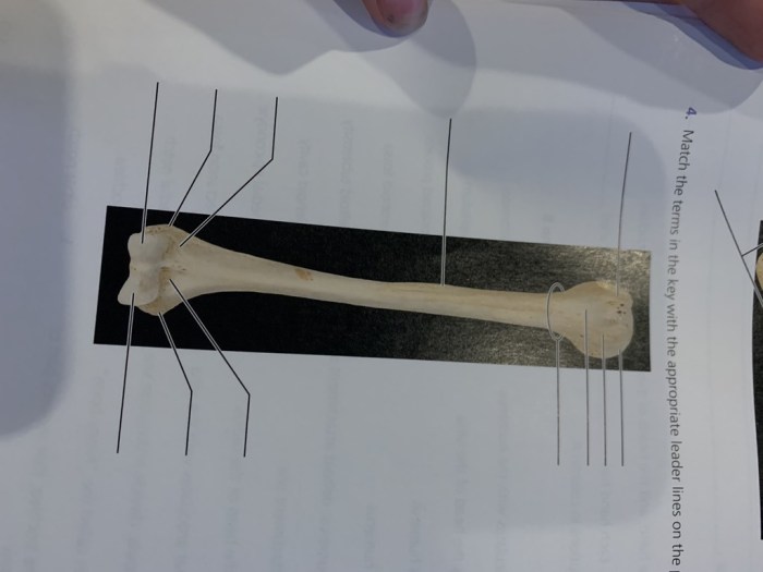
-*Humerus
The long bone of the upper arm, extending from the shoulder to the elbow.
-*Radius and Ulna
The two bones of the forearm, located side by side. The radius is on the thumb side of the forearm, while the ulna is on the little finger side.
-*Carpal Bones
Eight small bones that form the wrist.
-*Metacarpals
Five long bones that form the palm of the hand.
-*Phalanges
Fourteen bones that form the fingers and thumbs.
Joints of the Upper Limb
The joints of the upper limb allow for a variety of movements:
-
-*Shoulder Joint
A ball-and-socket joint that connects the humerus to the scapula (shoulder blade). It allows for a wide range of motion, including flexion, extension, abduction, adduction, and rotation.
-*Elbow Joint
A hinge joint that connects the humerus to the radius and ulna. It allows for flexion and extension of the forearm.
-*Wrist Joint
A condyloid joint that connects the radius and ulna to the carpal bones. It allows for flexion, extension, abduction, adduction, and circumduction of the hand.
-*Metacarpophalangeal Joints
Hinge joints that connect the metacarpals to the proximal phalanges (first finger bones). They allow for flexion and extension of the fingers and thumb.
-*Interphalangeal Joints
Hinge joints that connect the phalanges of the fingers and thumbs. They allow for flexion and extension of the fingers and thumbs.
Muscles of the Upper Limb
The muscles of the upper limb are responsible for moving the arm, forearm, and hand. These muscles can be divided into several groups based on their location and function:
-
-*Shoulder Muscles
These muscles surround the shoulder joint and are responsible for its movements, including the deltoids, supraspinatus, infraspinatus, teres minor, and subscapularis.
-*Elbow Flexors
These muscles are located on the anterior (front) side of the forearm and are responsible for flexing the elbow joint, including the biceps brachii, brachialis, and brachioradialis.
-*Elbow Extensors
These muscles are located on the posterior (back) side of the forearm and are responsible for extending the elbow joint, including the triceps brachii and anconeus.
-*Wrist Flexors
These muscles are located on the anterior side of the forearm and are responsible for flexing the wrist joint, including the flexor carpi radialis, flexor carpi ulnaris, and palmaris longus.
-*Wrist Extensors
These muscles are located on the posterior side of the forearm and are responsible for extending the wrist joint, including the extensor carpi radialis longus, extensor carpi radialis brevis, and extensor carpi ulnaris.
-*Hand Muscles
These muscles are located in the hand and are responsible for moving the fingers and thumb, including the flexor digitorum superficialis, flexor digitorum profundus, extensor digitorum, and abductor pollicis brevis.
The Pelvic Girdle

The pelvic girdle is a ring of bones that forms the lower part of the skeleton and connects the lower limbs to the axial skeleton. It consists of two hip bones (coxal bones) and the sacrum.
The pelvic girdle provides support and stability for the body, protects the pelvic organs, and allows for movement of the lower limbs.
Bones of the Pelvic Girdle
Each hip bone is formed by the fusion of three bones: the ilium, ischium, and pubis.
- The iliumis the largest and most superior part of the hip bone. It forms the upper part of the acetabulum, the socket into which the head of the femur fits.
- The ischiumis located inferior to the ilium. It forms the lower part of the acetabulum and provides attachment for the muscles of the buttocks.
- The pubisis located anterior to the ischium. It forms the anterior part of the acetabulum and provides attachment for the muscles of the abdomen.
Joints of the Pelvic Girdle, Exercise 9 the appendicular skeleton
The pelvic girdle is connected to the axial skeleton by the sacroiliac joints, which are formed between the sacrum and the ilium of each hip bone.
The hip bones are connected to each other by the pubic symphysis, which is a cartilaginous joint located at the anterior midline of the pelvis.
The Lower Limb
The lower limb, also known as the pelvic limb, is the part of the appendicular skeleton that extends from the hip joint to the toes. It consists of the thigh, leg, and foot. The thigh contains the femur, the longest bone in the body.
The leg contains the tibia and fibula, and the foot contains the tarsal, metatarsal, and phalangeal bones.The lower limb is responsible for supporting the weight of the body, allowing for locomotion, and providing stability. It is also involved in a variety of other functions, such as kicking, jumping, and squatting.
Bones of the Lower Limb
The lower limb consists of 30 bones, which are divided into three regions: the thigh, the leg, and the foot.Thigh
Femur
Leg
- Tibia
- Fibula
Foot
- Tarsal bones (7)
- Metatarsal bones (5)
- Phalanges (14)
Joints of the Lower Limb
The lower limb is made up of several joints that allow for a wide range of movement. These joints include:
- Hip joint
- Knee joint
- Ankle joint
- Subtalar joint
- Tarsometatarsal joints
- Metatarsophalangeal joints
- Interphalangeal joints
Muscles that Move the Lower Limb
The lower limb is moved by a variety of muscles, which are innervated by the sciatic nerve and its branches. These muscles can be divided into two groups:
-
-*Extensors
These muscles are responsible for extending the knee and ankle joints. They include the quadriceps femoris, gluteus maximus, and hamstrings.
-*Flexors
These muscles are responsible for flexing the knee and ankle joints. They include the gastrocnemius, soleus, and tibialis anterior.
Clinical Applications: Exercise 9 The Appendicular Skeleton

The appendicular skeleton is susceptible to various injuries and diseases, impacting its function and overall well-being. Understanding these clinical implications is crucial for healthcare professionals involved in diagnosis, treatment, and rehabilitation.
Common Injuries
Traumatic injuries to the appendicular skeleton are prevalent, ranging from fractures and dislocations to sprains and strains. Fractures, particularly in the long bones of the limbs, are often caused by high-impact forces or falls. Dislocations, on the other hand, involve the displacement of a bone from its normal joint position, typically resulting from sudden forceful movements.
Sprains and strains are injuries to ligaments and tendons, respectively, and can occur due to overexertion or sudden changes in direction.
Diseases Affecting the Appendicular Skeleton
Several diseases can affect the appendicular skeleton, leading to pain, deformity, and impaired function. Osteoarthritis, a degenerative joint disease, is a common condition that causes cartilage breakdown and inflammation in joints, often affecting the knees, hips, and spine. Rheumatoid arthritis is an autoimmune disorder that causes chronic inflammation and damage to joints, potentially affecting the appendicular skeleton.
Paget’s disease of bone is a metabolic disorder that leads to abnormal bone growth and remodeling, potentially affecting the long bones of the limbs.
Role in Rehabilitation
The appendicular skeleton plays a significant role in rehabilitation following injuries or surgeries. Physical therapy and rehabilitation programs aim to restore range of motion, strength, and function to the affected limbs. Exercises and therapies are designed to improve joint stability, muscle strength, and coordination.
In cases of fractures, immobilization techniques like casts or splints are used to promote bone healing and prevent further displacement. Rehabilitation focuses on gradually increasing load and movement to the injured area, allowing the appendicular skeleton to regain its optimal function.
Clarifying Questions
What is the function of the appendicular skeleton?
The appendicular skeleton provides support and protection for the limbs and girdles, facilitates movement, and allows for the attachment of muscles.
What bones make up the pectoral girdle?
The pectoral girdle consists of the clavicle and scapula.
What is the largest bone in the upper limb?
The humerus is the largest bone in the upper limb.
What is the function of the pelvic girdle?
The pelvic girdle supports the weight of the upper body, protects the pelvic organs, and provides attachment points for muscles.
